-
-
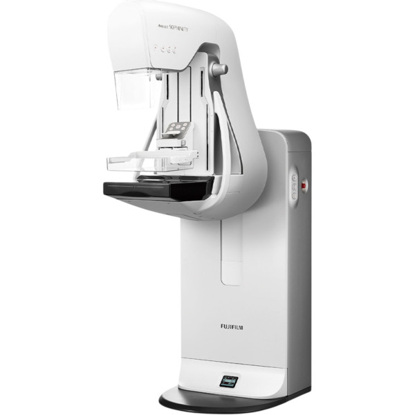 FUJIFILMMammography system AMULET SophinityMammography system AMULET SophinityRequest priceView details
-
-
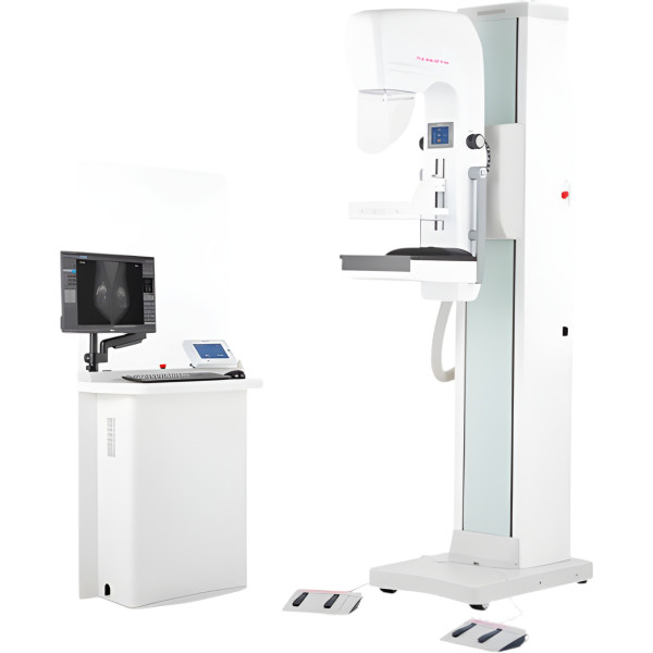 BEMEMSMammography system PINKVIEW-DR PRIMEMammography system PINKVIEW-DR PRIMERequest priceView details
-
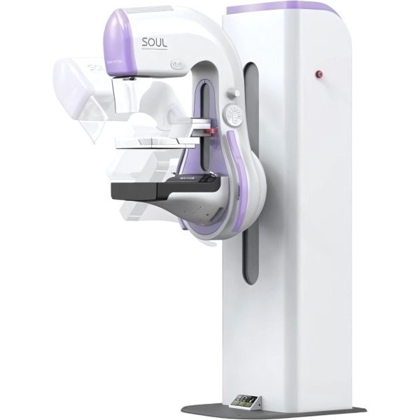 Medi-FutureMammography system SOUL DirectMammography system SOUL DirectRequest priceView details
-
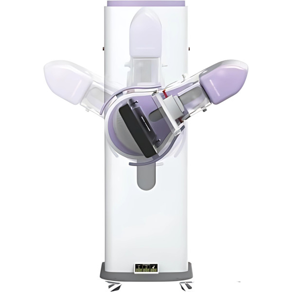 Medi-FutureMammography system SOUL IndirectMammography system SOUL IndirectRequest priceView details
Mammography systems
A mammography unit images the compressed breast with soft X-rays. The image shows masses and fine calcifications, which is why a facility relies on its mammography system for both screening and follow-up diagnostics. This section of the Finer catalog brings together digital mammography systems, and some of them offer tomosynthesis and biopsy. The manufacturers named in the product cards are FUJIFILM (Japan), Metaltronica, MEDI-FUTURE (South Korea) and ANGELL.
Why does a facility need a mammography system?
A screening program puts many women without symptoms through the room. There the unit is expected to deliver a fast exposure cycle and to be easy for the radiographer. A diagnostic center works differently. Patients arrive after an ultrasound or a breast specialist's exam, and the doctor needs spot views and tomosynthesis, sometimes a biopsy guided by the mammography unit. So a breast center usually has both a mammography system and an ultrasound scanner for breast imaging. Analog units with film or CR cassettes are still found in hospitals, but new purchases are mostly digital, because the image goes straight to the workstation and the archive.
Which mammography systems are in the Finer catalog?
For 2D work there is the SOUL Indirect mammography system, designed for screening and early diagnosis, and its SOUL Direct version for precise diagnostics. The PINKVIEW-DR PRIME has an a-Si detector with an inverter generator. The DM156 digital system works with a 24×31 cm detector, and the DM156 A sits next to it. Femaray-ME was made for dense breast tissue, while the AMULET Sophinity is presented as digital mammography with minimal radiation exposure.
For tomosynthesis, the Helianthus DBT mammography system supports both DBT and biopsy. There is also a mammography system with a 3D function and the Lilyum digital system. The product specs mention a 24×30 cm detector, a resolution of 6 line pairs per millimeter, biopsy in two projections, a 5 MP diagnostic monitor in the package and a technologist station. One configuration comes with two workstations at once.
How do you choose a mammography unit?
The detector determines most of what the doctor will see. A direct conversion detector (amorphous selenium) turns X-rays straight into an electric charge, while an indirect one (a scintillator over amorphous silicon, a-Si) first turns them into light. Both types are in clinical use. Compare actual figures: resolution and field size, then dose. A 24×30 cm field covers most of the breast in one exposure; with a smaller field, a large breast needs more exposures. A mammography unit with tomosynthesis costs more, and the radiologist reads dozens of slices instead of four standard views, so plan time for reporting.
Next, think about the daily routine of the room. Automatic exposure control and a choice of anode and filter for tissue density keep the dose in check, and a dose report is saved for each exam. Motorized compression with force control and a set of paddles for different projections matter beyond comfort: how the patient tolerates compression affects image quality. There are two workplaces, the technologist station at the unit and the reading station with 5 MP diagnostic monitors. The archive needs DICOM with transfer to PACS and the hospital information system. Tomosynthesis files take up far more space than 2D.
What does the price of a mammography system depend on?
A few models list a price in the card. Others are quoted through a commercial proposal. The cost is built from the detector and generator, the tomosynthesis license, the biopsy attachment, the number of workstations and monitors, and the image analysis software. A digital unit with tomosynthesis and biopsy costs noticeably more than a 2D screening system. To buy a mammography system without options you will not use, tell us whether it is for screening or diagnostics, how many exams a day you expect, what room you have and whether your facility runs PACS.
Where to buy a mammography system?
Finer Ukraine LLC has supplied medical facilities since 2015 and provides mammography systems to hospitals and to diagnostic or breast centers. Finer's doctors and engineers match the model to your patient flow and the tasks of the room, then prepare the commercial proposal and tender documents. The team delivers and installs the unit, then commissions it. Radiographers and doctors are trained to work with it. If you are opening a center from scratch, see our turnkey solutions. General radiography is covered by the X-ray units, and the rest of the equipment is in the X-ray machines section. Questions about a specific system go to 0 800 334 585 or through the form on our contacts page.
FAQ
1. What is tomosynthesis in mammography?
The tube sweeps in an arc over the breast and takes a series of low-dose images, and the software builds slice images from them. Tissue overlaps less than on a standard 2D image, which shows especially in dense breasts. The Helianthus DBT from Metaltronica is one of the units that can do it.
2. How is a digital mammography unit different from an analog one?
A digital unit sends the image straight to the workstation without cassettes or processing, and the image is easy to share with colleagues or store in PACS. An analog unit needs film and cassettes plus a processor, and the image takes longer to reach the doctor. The Finer catalog lists digital systems.
3. What does a mammography room need?
A mammography unit is a source of ionizing radiation. The room needs radiation shielding built to a design project. It also needs dosimetry monitoring and a license to use ionizing radiation sources, plus a stable power supply of the right capacity. A digital unit also needs a technologist station and a reading workstation with a diagnostic monitor.
4. Mammography or breast ultrasound: which does a center need?
Usually both. Mammography shows fine calcifications well, while ultrasound helps assess a mass in dense tissue and tell a cyst from a solid lesion. Patients are often sent for mammography after ultrasound or vice versa, so breast centers keep both methods under one roof.
5. Who operates a mammography unit?
A radiographer takes the images and a radiologist reports them. The radiographer works at the station next to the unit, and the doctor reviews images on a workstation with 5 MP diagnostic monitors. If several doctors report, you will need more reading stations.
Categories by purpose



- Solutions
- Finer Ukraine