-
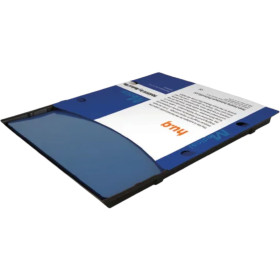
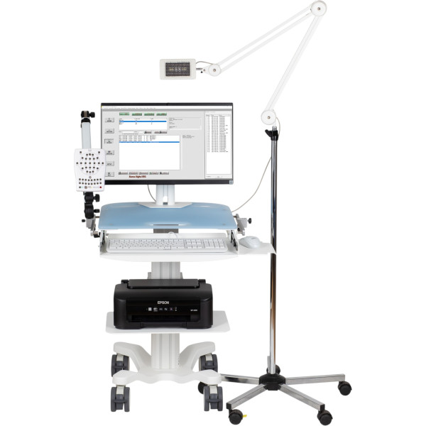 EMS Biomedical32-channel electroencephalograph Sienna Ultimate32-channel electroencephalograph Sienna UltimateRequest priceView details
-
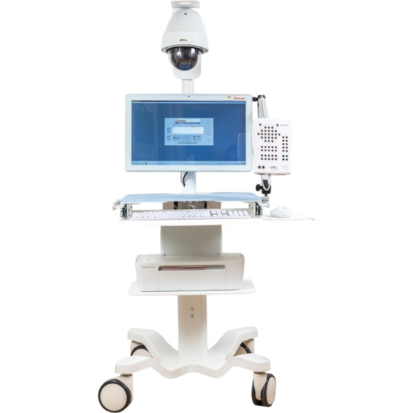 EMS Biomedical40-channel electroencephalograph Sienna40-channel electroencephalograph SiennaRequest priceView details
-
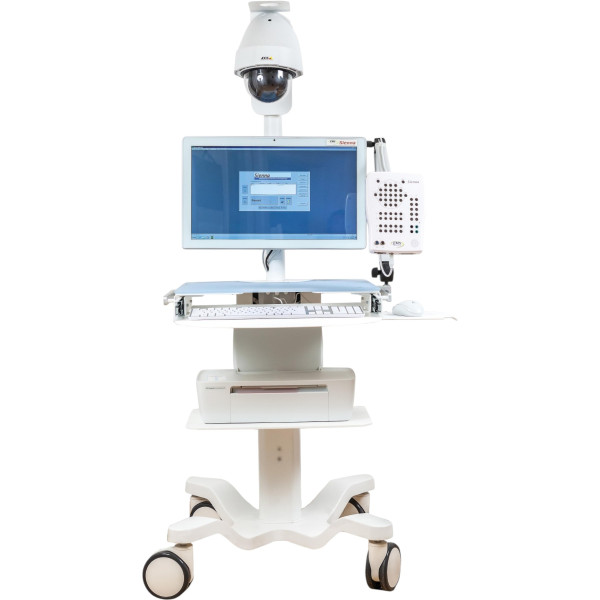 EMS Biomedical64-channel electroencephalograph Sienna64-channel electroencephalograph SiennaRequest priceView details
Electroencephalographs (EEG)
An EEG machine records the electrical activity of the brain through electrodes on the scalp. For a neurology department or a functional diagnostics room it is an everyday device. Channels and modules are therefore chosen to fit the room's tasks. This section of the Finer catalog lists Sienna EEG systems by the Austrian company EMS Biomedical with 32, 40 or 64 channels for routine, long-term and high-density recordings.
What is an EEG machine for?
Electroencephalography remains the main instrumental method in diagnosing epilepsy. It is also used in disorders of consciousness and sleep, after traumatic brain injury and for monitoring in intensive care, when the patient is unconscious and the doctor has few other ways to assess brain function at the bedside. A routine recording is short. Epileptologists, though, need recordings that last for hours with synchronized video, because only then can they match what they see on screen during a seizure with what the EEG trace shows at that same second.
How do you choose an EEG machine?
Start with the channels. A routine EEG on the 10-20 system needs 19 to 21 leads. For video-EEG, 32 to 40 channels are convenient, and 64 or more are taken for dense electrode layouts. Next comes recording length: if the room will see epilepsy patients, long recordings with video are needed. Caps speed up electrode placement, while individual cup electrodes give flexibility. In the software, look at montages and filters, event markers, plus export in a format your hospital information system accepts. The device itself needs a quiet room with proper grounding, away from electrical noise.
Which EEG systems are in the Finer catalog?
The Sienna Ultimate 32-channel EEG is designed for routine and long-term recordings. The Sienna 40-channel system is modular, so it can be expanded. The Sienna 64-channel system is modular too and is chosen when many leads are needed. For cardiology there are electrocardiographs, and other functional diagnostics equipment is in the Diagnostic Equipment section.
Where to buy an EEG machine?
Finer Ukraine LLC has supplied equipment to medical facilities since 2015, and EEG systems for neurology are part of that range. To buy an EEG machine without modules you will never use, tell us which studies the room will run, and we will prepare a quote for that configuration. The doctors and engineers on our team take care of the tender paperwork and delivery, then set up the system and train the staff. Our number is 0 800 334 585, and there is also a contacts page.
FAQ
1. How does an EEG machine work?
Electrodes on the scalp pick up the electrical potentials of the brain cortex. They are very weak. An amplifier boosts the signal, and the software shows it as a set of traces for each lead. The doctor assesses the rhythms and their symmetry along with any abnormal waves, including during hyperventilation or light stimulation tests.
2. What does an EEG show?
How the brain works at the time of recording: the main rhythms, how they change in sleep and during tests, plus epileptiform activity. That is why EEG is the main method in diagnosing epilepsy. It is also used in disorders of consciousness, after brain injury and for monitoring in intensive care.
3. What is video-EEG monitoring?
A long EEG recording synchronized with video of the patient. The doctor sees what happened to the person at the moment the trace changed, and this is how an epileptic seizure is told apart from other conditions. Systems with 32 to 40 channels, such as Sienna Ultimate or the 40-channel Sienna, suit video-EEG.
4. How does EEG differ from MRI?
They are different methods. EEG shows the electrical work of the brain in real time and catches epileptic activity. MRI shows brain structure, such as tumours, the after-effects of a stroke or developmental anomalies. They are often ordered together, and the doctor chooses the method.
5. Cap or individual electrodes for EEG?
Caps with electrodes on the 10-20 system speed up placement, so they suit a steady flow of routine recordings. Individual cup electrodes give flexibility in positioning and are convenient for long recordings. Both need electrode gel or paste, which should be budgeted as consumables.
Categories by purpose

Recently viewed

- Solutions
- Finer Ukraine